tree in bud lesion
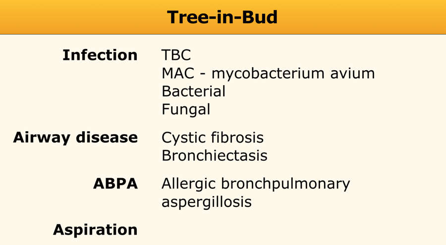
In clinical practice the tree-in-bud pattern may be indicative of a wide range of pathogens including. Simply put the tree-in-bud pattern can be seen with two main sites of disease 3.
Tree In Bud Pattern Pulmonary Tb Eurorad
The Tree-in-bud Pattern.
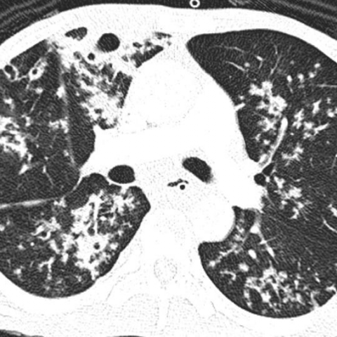
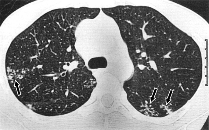
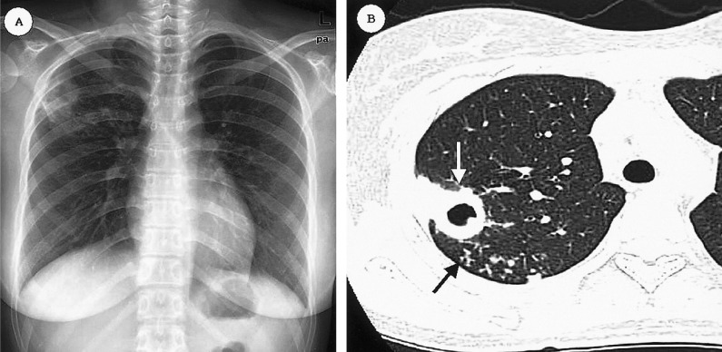
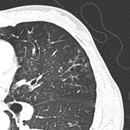
. Usually somewhat nodular in appearance the tree-in-bud pattern is generally most pronounced in the lung periphery and associated with abnormalities of the larger airways. Post-mortem radiograph of patient with active pulmonary tuberculosis demonstrating tree-in-bud lesion boxed area with smooth marginated bronchiole tree and. The tree-in-bud sign is a nonspecific imaging finding that implies impaction within bronchioles the smallest airway passages in the lung.
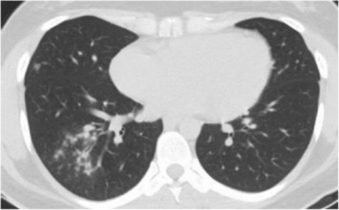
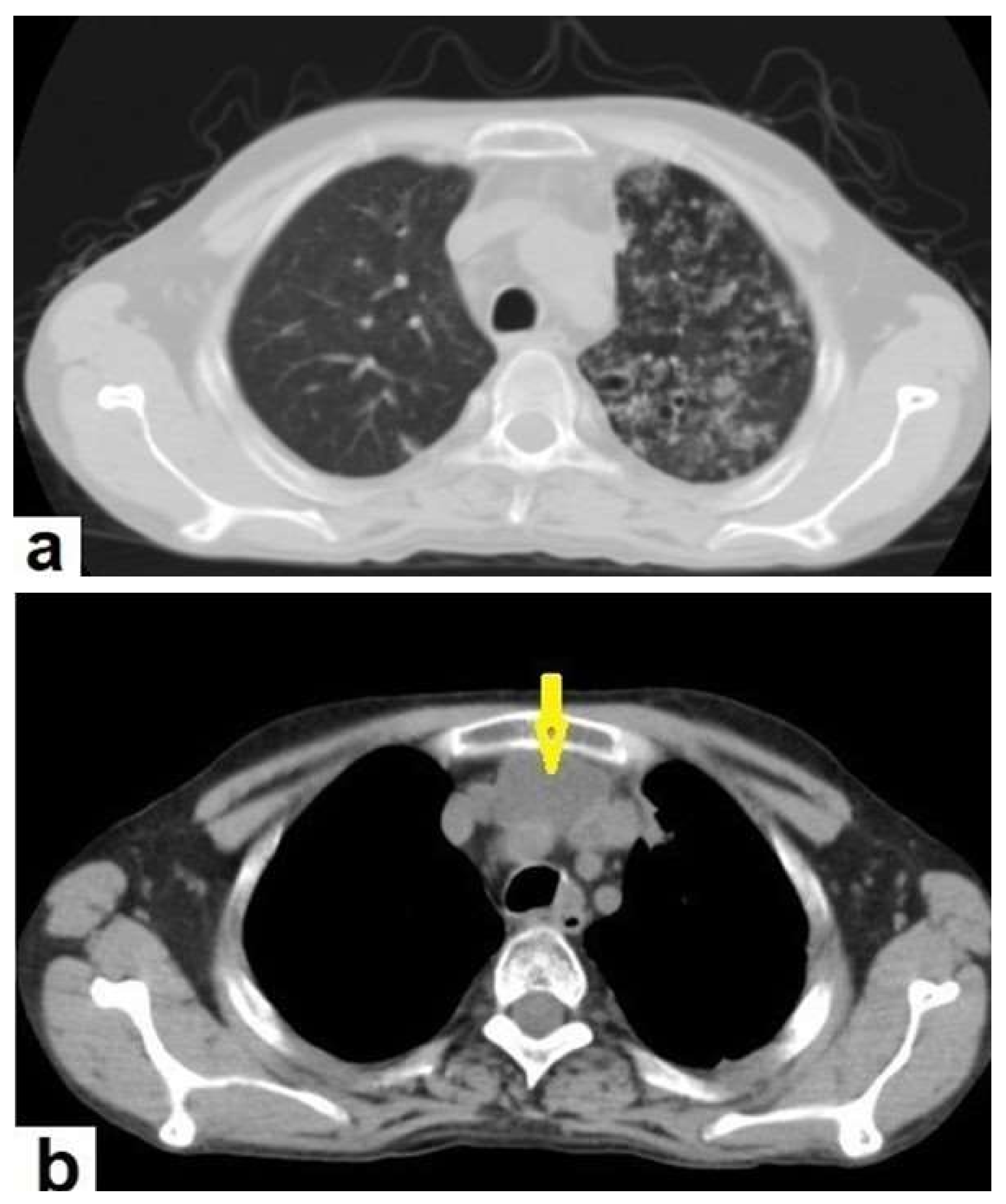
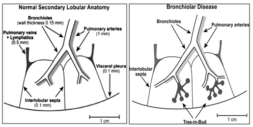
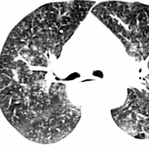
The tree-in-bud pattern represents centrilobular branching structures that resemble a budding tree. Centrilobular nodules with a linear branching pattern are consistent with tree-in-bud appearance in a patient with endobronchial spreading of post-primary tuberculosis. The tree-in-bud-pattern of images on thin-section lung CT is defined by centrilobular branching structures that resemble a budding tree.
The tree-in-bud sign is a nonspecific imaging finding that implies impaction within bronchioles the smallest airway passages in the lung. A Common Finding On Thoracic Ct Scans. We investigated the pathological basis of.
In the 26 patients. Distal pulmonary vasculature More specifically the pattern can be manifest because of the following disease processes often in combination. The tree-in-bud pattern is commonly observed in CT of the lungs because it consists of small centrilobular nodules of soft tissue attenuation connected to multiple.
The pattern reflects a spectrum of endo- and peribronchiolar disorders including mucoid. By Matt Oct 16 2022 Mast Trees. Tree-in-bud refers to small airway at the bronchiole level involvement of lesions resulting in expansion of the airway and infiltration of pathological substances into the tube.
At examination with CT centrilobular lesions nodules or branching linear structures 2-4 mm in diameter were most commonly seen n 39 95. The peculiarity of the case was that there were streaky areas of enhancement around the lesion in the brain parenchyma which resembled tree-in-bud like appearance. Distal airways more common 2.
Bronchiolesfilled with pus or infla See more. 87 rows mid-bronchial lesion. 87 rows The tree-in-bud sign indicates bronchiolar luminal impaction with mucus pus or.
Non-infectious causes of the tree-in-bud sign include diffuse panbronchiolitis cystic fibrosis immotile cilia syndrome and congenital immunodeficiency states. Tree-in-bud sign refers to the condition in which small centrilobular nodules less than 10 mm in diameter are associated with centrilobular branching nodular structures 1 Fig. The tree-in-bud sign is a radiographic appearance seen on chest x-rays and CT scans that is indicative of pulmonary nodules or mass.
Clustered Micronodules As Predominant Manifestation On Ct A Sign Of Active But Indolently Evolving Pulmonary Tuberculosis Plos One

Kjr Korean Journal Of Radiology
The Radiology Assistant Hrct Basic Interpretation
Diagnostics Free Full Text The Diagnostic Deceiver Radiological Pictorial Review Of Tuberculosis Html
Koreamed Synapse
Patterns Of Lung Disease Springerlink
The Korean Journal Of Internal Medicine
Epos

Areas Showing A Mosaic Pattern Of Attenuation And Tree In Bud Opacities Download Scientific Diagram

Tree In Bud Pattern Semantic Scholar

Tree In Bud Pattern Semantic Scholar
Tree In Bud Sign Lung Radiology Reference Article Radiopaedia Org
Covid 19 Pneumonia A Pictorial Review Of Ct Findings And Differential Diagnosis Egyptian Journal Of Radiology And Nuclear Medicine Full Text
Tree In Bud Sign Lungs
It Is Not Always Tuberculosis Tree In Bud Opacities Leading To A Diagnosis Of Sarcoid Shm Abstracts Society Of Hospital Medicine

Tree In Bud Sign An Overview Sciencedirect Topics
It Is Not Always Tuberculosis Tree In Bud Opacities Leading To A Diagnosis Of Sarcoid Shm Abstracts Society Of Hospital Medicine

Tree In Bud Pattern At Thin Section Ct Of The Lungs Radiologic Pathologic Overview Radiographics
Low Pectoralis Muscle Index Cavitary Nodule Or Mass And Segmental To Lobar Consolidation As Predictors Of Primary Multidrug Resistant Tuberculosis A Comparison With Primary Drug Sensitive Tuberculosis Plos One